
Introduction to Electrophoresis:
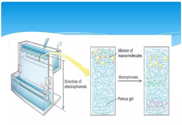
The movement of charged particles under the action of an electric field towards an electrode opposite to their electrical properties is called electrophoresis (EP). The technique of utilizing charged particles moving at different speeds in an electric field to achieve separation is called electrophoresis.
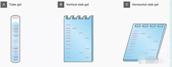
Nucleic acid electrophoresis is an important tool for nucleic acid research and is an integral part of techniques such as nucleic acid probes, nucleic acid amplification and sequence analysis. Nucleic acid electrophoresis is usually carried out in agarose gels or polyacrylamide gels. Agarose and polyacrylamide with different concentrations can form gels with different sizes of molecular sieve mesh, which can be used to separate nucleic acid fragments of different molecular weights.
Principle of electrophoresis:
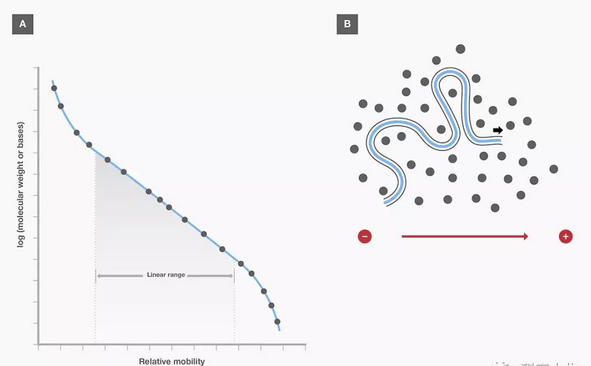
It is mainly utilized that DNA molecules have a charge effect and a molecular sieve effect when swimming in agarose gels. DNA molecules are negatively charged in a pH solution above the isoelectric point and move toward the positive pole in an electric field. Due to the structurally repetitive nature of the sugar-phosphate backbone, the same amount of double-stranded DNA has an almost equal net charge, so they can move toward the positive pole at the same rate.
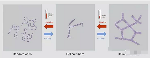
Agarose gels have a network structure, and molecules of matter are resisted as they pass through, and large molecules are resisted in surges, so that in gel electrophoresis, the separation of charged particles depends not only on the nature and amount of net charge, but also on the size of the molecules, and so it becomes possible to use it for the separation of nucleic acid fragments of different molecular weights.
lit. descending dimensional narrative:
Imagine fragments of nucleic acids of different molecular weights as fat and thin people with no obstacles since the net charges of the equal amounts are all the same. The time to run to the positive terminal is equal.
Now we add the barrier (agarose gel) network structure. The skinny guy passes through the net faster than the fat guy passes through the time. So it results in the skinny guy running faster and getting to the finish line first. People of the same size all have almost the same rate.
So I arrange for a couple of weighed people to join me in an obstacle course run. It is then possible to estimate the weight in the area of the crowd. That is, the molecular weight of the nucleic acid, which is also known as the length of the so-called DNA strand due to the structurally repetitive nature of the sugar-phosphate backbone.
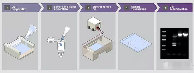
Nucleic acid electrophoresis process steps
- Selection and preparation of gels
Agarose and polyacrylamide are the two most commonly used gel matrices for nucleic acid separation. Both materials are three-dimensional matrices with pore sizes suitable for nucleic acid separation and are non-reactive with the sample. The pore size can be adjusted by changing the percentage of matrix to effectively separate nucleic acids of different sizes.
- Preparation of standards and samples
- a. Nucleic Acid Standard Selection
When running gels, reference samples containing nucleic acids of known size are often referred to as standards, markers, or molecular weight standards, and are used for estimating the size of the intended sample. - b. Sample and standard preparation
The amount of DNA to be sampled into the gel must be calculated to ensure that the target bands are well separated and used for visualization and detection. Although the use of fluorescent dyes is sufficient to detect 1 – 100 ng/band of DNA, the minimum detectable amount depends on the dye used. Note that overloading of samples or standards can cause band smudging and obscuring of nearby bands, resulting in poorly resolved bands, especially when fragments are similar in size. - c. Sampling dye and buffer selection
Gel electrophoresis is prepared with a gel upload buffer (usually a 6X or 10X stock solution) added to the sample.
- Running electrophoresis
Electrophoresis is performed after gel, standards and samples have been prepared. The gel must be completely solidified before removing the comb and adding electrophoresis buffer. The gel comb should be lifted up smoothly to avoid tearing the gel and distorting the gel holes. After removing the comb and adding buffer, take care to remove any air bubbles from the wells. For polyacrylamide gels, the gel holes should be thoroughly rinsed with buffer to remove residual unpolymerized acrylamide.
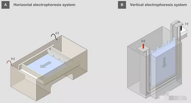
Horizontal gels should be oriented toward the gel box with the sample wells on the negative side to allow the sample to be moved to the positive side after electrophoresis is initiated. This orientation can be noted as “run to red” since the positive electrode is usually red. In vertical gel cassettes, the holes are designed to be at the top.
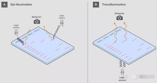
- Visualizing the sample in the gel
After the gel run is complete, the sample needs to be visualized and analyzed. Since nucleic acids are not visible under normal lighting, a visualization assay is needed. - Recording the gel
After visualization, the nucleic acid gel is usually archived for recording and analyzing the electrophoresis results.
Source: https://www.sperikon.com/1225.html